






Precision Approach to Viral Immunotherapy
Researchers have developed a method using synthetic super-enhancers to enable precision viral immunotherapy, representing an advancement in targeted therapeutic approaches. This technique focuses on enhancing the specificity and effectiveness of viral vectors used in treatment applications.
The development addresses the need for more precise delivery systems in immunotherapy contexts. The approach involves creating custom molecular tools for efficient genetic manipulation.
Molecular Cloning Techniques
A specific custom destination vector was built for efficient Golden Gate cloning of enhancer fragments, facilitating the assembly of synthetic super-enhancers. This cloning method allows researchers to precisely control the genetic components used in therapeutic development.
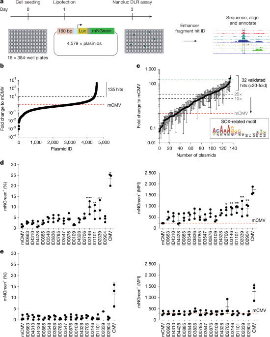
For reporter gene expression, the reporter gene cassette NanoLuc-Ires-mNGreen-pA was placed downstream of a mCMV promoter. This configuration enables monitoring of gene expression and therapeutic activity in experimental systems.
Molecular Techniques and Protein Expression
The research employed precise molecular biology techniques for DNA manipulation and analysis. PCR purification used Agencourt AMPure XP magnetic beads from Beckman Coulter according to manufacturer instructions, ensuring clean DNA samples for downstream applications.
For quantification of PCR products, researchers used Tapestation and reagents according to manufacturer instructions. This provides accurate measurement of DNA concentrations and quality.
Protein Production
Protein expression involved specific human transcription factors relevant to the research. His-tagged human SOX2 and SOX9 were expressed from a pET28a backbone cloned by G. Roberts in the Soufi Laboratory.
The expression system utilized Rosetta 2(DE3) pLys competent cells from Novagen. This bacterial strain is designed for efficient protein expression, particularly for challenging proteins. The source did not provide details about specific expression yields or purification methods.
Tissue Testing and Viral Delivery
Experimental testing involved precise viral delivery to tissue slices. A 5 µl volume of virus was pipetted onto the centre of each tissue slice using a 10 µl tip without touching the tissue. This careful technique minimizes tissue damage and ensures consistent viral application.
Researchers administered three repeat doses of 5 µl virus to each slice at 5-minute intervals. This staggered delivery approach allows for better tissue penetration and distribution. In total, 20 µl of virus was added per slice, providing a standardized treatment volume.
Following viral administration, slices were incubated for 7 days to allow for therapeutic effects to develop. The medium was replaced on day 3 or 4 to maintain tissue health and remove waste products.
Data Analysis and Visualization Tools
Comprehensive data analysis employed multiple software platforms. Data analysis was performed using:
- Microsoft Excel v.16.23 for Mac
- GraphPad Prism v7
- RStudio v1.1.456
These tools provide statistical analysis, graphing capabilities, and programming environments for thorough data examination.
Statistical and Visual Representation
For statistical representation, error bars are shown as the standard deviation (s.d.) of the mean. This measurement indicates the variability within experimental data sets. The source did not provide details about specific statistical tests or significance thresholds used.
Visualization and illustration utilized specialized software tools. For illustrations, BioRender and Adobe Illustrator 22.0.1 were used to create clear scientific figures and diagrams.
Additional computational analysis employed open-source bioinformatics programmes including:
- bedtools
- GREAT
- fastasplitter
- HOMER
- MEME
These tools support genomic data processing, motif discovery, and functional annotation tasks.
In Vivo Application and Tumor Treatment
The research extended to in vivo applications using specific viral constructs. AAV1-SSE-7-mCMV–HSV-TK-V5–mCherry was injected at around 3.58 × 10^14 viral genomes per ml and 7.2 × 10^13 viral genomes per ml in PBS. These concentrations represent different treatment doses for experimental evaluation.
AAV was injected into tumours as described above, applying the synthetic super-enhancer approach to cancer models. After 7 days, tumours were imaged using a stereomicroscope and surgically excised.
Further information on research design is available in the Nature Portfolio Reporting Summary linked to this article. Readers interested in methodological details can consult this additional resource. The source did not provide details about specific therapeutic outcomes or efficacy measurements.
As with any emerging therapeutic approach, individuals should consult a healthcare professional for personalized medical advice. This research represents laboratory development that requires further validation before clinical application.
Frequently Asked Questions
How were synthetic super-enhancers cloned for precision viral immunotherapy?
A specific custom destination vector was built for efficient Golden Gate cloning of enhancer fragments, and the reporter gene cassette NanoLuc-Ires-mNGreen-pA was placed downstream of a mCMV promoter.
What experimental methods were used for virus delivery in tissue slices?
A 5 µl volume of virus was pipetted onto the center of each tissue slice without touching it, with three repeat doses added at 5-minute intervals totaling 20 µl per slice, followed by 7-day incubation with medium replacement on day 3 or 4.
What viral concentration was used for AAV1-SSE-7-mCMV–HSV-TK-V5–mCherry injections in tumors?
AAV1-SSE-7-mCMV–HSV-TK-V5–mCherry was injected at approximately 3.58 × 10^14 viral genomes per ml and 7.2 × 10^13 viral genomes per ml in PBS, with tumors imaged and excised after 7 days.